Periapical Cyst Treatment
Periapical Cyst Treatment
What is a Periapical Cyst?
Also known as a radicular cyst, the periapical cyst is a common occurrence in the jaw. Necrosis of the pulp occurs as a result of infection of the tooth. Due to toxins emerging from the apex of the tooth periapical inflammation settles in. The Malassez epithelial rests are stimulated in the periodontal ligament causing the formation of a periapical granuloma. Blood supply is arrested in the epithelium and a necrosis develops. The lesions are incidentally discovered on radiographs more often. A periapical cyst develops between 20 years and 60 years of age with the maxilla being affected more than the mandible.
Clinical features of Periapical Cyst
The periapical cyst is progressive and forms slowly. It is usually asymptomatic but can cause pain secondary to infection. There is swelling, which expands rapidly due to inflammatory edema. As it advances, part of the mouth wall is resorbed and a soft fluctuant swelling bluish in color beneath the mucous membrane is formed. The bone has the thickness of an egg shell and upon pressure a crackling sensation is felt.
What are the Causes of Oral Lichen planus?
The radicular cyst pathogenesis is divided into three phases:
Initiation: Epithelial cell rests of Malassez respond to stimulation due to bacterial infection of the pulp and as a response to necrotic pulp tissue.
Formation: The proliferating epithelium lines the central cavity in the periapical area. The epithelial mass increases in size with the peripherial division of cells and the cells from the mid portion of the mass are separated from nutrition. The epithelium-lined cavity is filled with fluid due to liquefactive necrosis of the innermost cells.
Enlargement or expansion: The innermost cellular debris raises the lumen of the cyst increasing the size of the cyst. Gamma globulin levels are found in the fluids of radicular cysts.
Radiologic evaluation
Ovoid to round radiolucency is noticed with a narrow, opaque margin. The margin is indicative of rapid enlargement of the cyst and is contiguous with the lamina dura. Most cysts are < 1.5 cm in diameter and root resorption of the offending tooth and the adjacent teeth are noticed in long-standing periapical cysts.
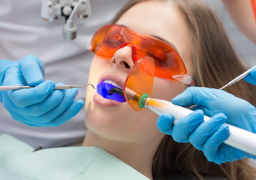
At Dental Solutions, cyst enucleation is performed with the use of conventional laser techniques. Research indicates that Nd:YAG and Er:YAG lasers can successfully eliminate the E.Faecalis and Escherichia coli thus making laser the most appropriate device for the treatment of periapical cysts. Some efficacious wavelengths that are used: ER:YAG 2940 nm and Nd:YAG 1064 nm diffuse dentin infections of varied thicknesses. The microbial population in the canals is removed with the maximum effect when the laser is combined with sodium hypochlorite in ideal concentrations for irrigation. Laser penetrates areas of the secondary canal where irrigation solutions cannot reach